PLATFORME
Presentation
Located on the campus of the University of Toulouse Paul Sabatier, the Light Iimaging Toulouse CBI platform has a wide range of photonic imaging technologies available. It provides solutions to a wide range of biologics questions, from developmental biology to microbial genetics.
Certified to ISO 9001 and NFX50-900 standards, LITC is part of the GIS Genotoul and is attached to the Toulouse Réseau Imagerie (TRI). Access to its facilities is open to internal and external public research teams, as well as to private companies.
LITC is also involved in research and development on a new imaging concept, using coherent light based on random optical scribes known as RIM (Random Illumination Microscopy), which overcomes the limitations of existing commercial systems to image molecular phenomena on living biological tissue at 100nm resolution.
The development priorities address the biological issues explored by the CBI’s research teams, with a strong interdisciplinary component including:
– Single molecule analysis (in interaction with the METi platform for electronic microscopy)
– New super-resolution techniques in structured and random illumination (SIM, 3D-SIM, RIM) and localization or ‘pointillist’ microscopy (localization microscopy: PALM, STORM, PAINT, sptPALM)
– Correlative Light Electron Microscopy (CLEM) – in interaction with the METi platform for electronic microscopy
– Opto-manipulation techniques (optical tweezers and opto-genetics module coupled to a spinning disc)
Services: Training, advice and expertise
– Support and collaboration on research projects
– Training in the use of available technologies
– Help in optimising protocols or acquisitions
– Assistance with analysis
– Participation in continuing education (CNRS Formation Entreprise, Doctoral School, University, etc.)
Nikon live
Specifically dedicated to video microscopy, this microscope is equipped with a Hamamatsu OrcaFlashV2 camera, a Lumencor ® 7-colour excitation source, multi-band dichroic mirrors and an external filter wheel for rapid multi-colour acquisitions to image fluorophores such as DAPI, CFP, YFP, GFP, mCherry, Cy3 and Cy5. The presence of a temperature-controlled chamber and the PFS focus maintenance system means that live samples can be imaged over time.
Objectives available are a 20x/0.8 Mimm, a 40x/1.3 oil, a 100x/1.4 oil, a 100x/1.3 oil Ph3 and a 20x/0.5 sec long working distance.
Leica Spinning disc coupled with an optogenetic system
Spinning disk produces high-speed multidimensional images with optical sectioning, high SNR and minimal phototoxicity. Specifically dedicated to live imaging with high temporal resolution and low phototoxicity, this inverted Leica spinning-disk is equipped with 4 excitation diodes laser at 405 nm, 488 nm, 561 nm and 637 nm ( 100 – 150 mW) and classical emission filters to image the most commonly used fluorophores (DAPI, eGFP, mcherry, mKate, Alexa647). The disk is a Yokogawa CSU-X1, the camera is a CMOS (Hamamatsu OrcaFlash4 V2+). It is also equipped with a temperature-controlled chamber allowing long time imaging and a 250 micron piezo for rapid z-control. The acquisition software is MetaMorph 7.10.
This microscope also allows optogenetic manipulations. It is equipped with a Lego IR system. The 1470 nm laser (1 W) allows local heating of the sample for spatial and temporal control of protein expression (under Hsp70 promoter). It is also equipped with a 457 nm laser diode under the control of galvanometric mirrors, allowing localized activation of photosensitive proteins.
Leica equipped with optical tweezers
A mutlimodal imaging system that combines rapid 3D fluorescence imaging with remote focusing, and a versatile optical tweezer system compatible with quantitative force measurement by interferometry on living tissue. Force measuments, and mirorheology are possible with the sysem, also in living tissues.
The Home made system is a combination of a fluorescence microscope and an active optical trapping system. This trapping system comprises two components, one to precisely control the position of the optical trap on the sample and a back focal plane interferometry (BFPi) system to track the position of the trapped object relative to the laser centre. The laser used for trapping was centred at 1064 nm (Fiber laser 1W). For the calibration and measurements, a piezoelectric mirror conjugated to the pupil plane of the microscope (Thorlabs FSM75-P01) is used to obtain a nanometric deflection of the laser. A 3-axis piezoelectric stage (Piezo concept BIO3) is also used to focus precisely on the trapped object. The optical trapping system is implemented in a conventional inverted fluorescence microscope (Leica DMI6000 B). The objective used is a 100x magnification objective with a numerical aperture of 1.4 (Leica HCX PL APO). To optimise the fill factor of the infrared laser to 85% of the objective pupil plane, an afocal X4 telescope consisting of two relay lenses was used (Thorlabs ACA254-050-1064 and ACA254-200-1064).
The microscope is optimised for simultaneous imaging of GFP and RFP in combination with the infrared laser trap using a 3-band dichroic (Semrock Di03-R405/488/561/635-t1-25×36). Imaging and optical clamps are achieved by combining a National Intruments acquisition card with the Inscoper synchronisation box. The Inscoper software manages all the synchronisation steps.
Nikon equipped with Impetux commercial optical tweezer
A multimodal imaging system that combines 2D fluorescence and direct force measurements with optical tweezers based on momentum measurements with special optics for back-focus interferometry. Multiple trapping (up to 256) is possible. Force measurements, microrheology, constant force measurements are possible with this system for living cell and in vitro experiments. impetux SA’s system is a combination of a fluorescence microscope and an active optical trapping system.
This trapping system consists of two components, one for the precise control of the position of the 25KHZ optical trap on the sample thanks to AOD (Acousto Optical Device) and a direct force measurement instrument based on back focal plane interferometry (BFPi) (oil immersion Imputex Sensocell detector NA=1.2) relative to the centre of the 25KHZ laser.
The optical trapping system is implemented in a conventional widefield inverted fluorescence microscope (Nikon TiE2) with perfect focus. The objective used is a water X60 magnification objective with a numerical aperture of 1.2 (Nikon Plan APO VC X60/.1.2 WI OFN2 DIC N2). Imputex software manages all the synchronisation steps with Micromanager.
2 microscopes Leica SP8
Specifically dedicated to the acquisition of 3D samples, the 2 Leica SP8 systems (one upright and one inverted) are equipped with 2 photomultipliers and a high-sensitivity hybrid detector.
They are equipped
Objectives available are 20x/0.75 imm, 40x/1.3 oil and 63x/1.4 oil. The right-hand stand is also equipped with a 25x/0.95 water immersion long working distance physiological objective.
2 microscopes Zeiss + Airyscan
Specifically dedicated to the acquisition of 3D samples, the 2 Zeiss inverted systems (one 710 and one 880) are equipped with 2 photomultipliers, 2 high-sensitivity GasP detectors and an Airyscan module on the Zeiss 880 to increase resolution. Equipped with temperature-controlled chambers, they can also image living samples over time.
They are equipped with a 405nm laser diode and lasers at 488, 561nm and 633nm. The Zeiss 710 also has excitations at 458nm and 514nm.
Objectives available are a 20x/0.8 sec, a 25x/0.8 imm long-distance, a 40x/1.3 oil and a 63x/1.4 oil.
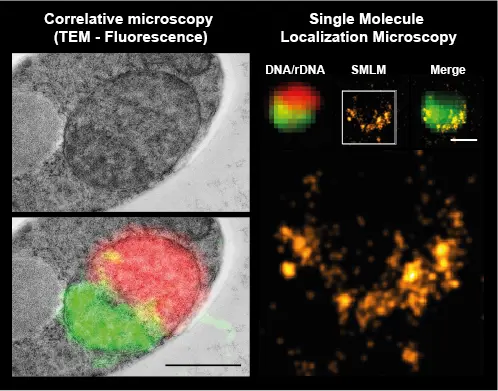
Nikon I-SPT microscope for 3D single molecule imaging (PALM, STORM, PAINT, sptPALM)
This microscope is dedicated to single molecule localization microscopy (SMLM) and allows PALM, STORM, PAINT or spt-PALM (single particle tracking on living cells) acquisitions.
This Nikon TiE microscope is equipped with an EM-CCD camera (Ixon ULTRA DU897 Andor – 512*512 pixels – pixel size: 16µm before magnification), a TIRF 100X oil NA 1.49 DIC, a motorised stage, a Mad City Lab Nano Z100 TI piezometric insert, a STORM quadriband dchroic cube, an external emission filter wheel (Sutter – Lambda 10-B) with the following filters: 536/40; 624/40; QUAD; 525/50; 600/50; 705/72.
Illumination is provided by a Nikon LU4 laser bench that can inject the following 4 lasers separately or simultaneously:
561nm at 200mW (Sapphire – Coherent)
488nm at 200mW (Sapphire – Coherent)
405nm at 100mW (Coherent)
647nm at 300mW
The camera and lasers are synchronised via the ENS HW BOX.
The system is temperature-stabilised, mounted on an active optical table and the instrumental drift is corrected via a Z-focus maintenance system (PFS). Drift in the X and Y axes requires post-acquisition correction.
A deformable mirror (MicAO, Imagine Optics) is used to modulate the system’s PSF according to the Zernike polynomial and to introduce anisotropic aberrations for the 3D localisation of single molecules (astigmatism, tetrapod).
Offset phase rings and an additional camera enable phase-contrast imaging to be carried out using the same lens as that used for single-molecule detection, making it possible to co-locate phase imaging and super-resolved SMLM imaging. This is particularly useful for studying bacteria.
2 microscopes Nikon RIM
Random Illumination Microscopy is a super resolution technology, high speed and low photo damages.
On both systems, a spatial light modulator (SLM) allows us to design various sequences illumination patterns. Consequently, these systems can be used for RIM imaging as well as SIM, ISM, TIRF, etc.
RIM1: dedicated to microbiology, fixed cells and live cells in culture is equipped with 2 cameras (Orca-Fusion) for 2 simultaneous color acquisitions. Oxxius 6 colours lasers are available (405nm, 458nm, 488nm, 515nm, 561nm, 638nm) and the PSF (perfect focus system) and temperature-controlled chamber permit to image living samples over time.
100x/1.49 APO SR TIRF oil object is dedicated to this system.
RIM2: dedicated to 3D models and thick samples is equipped with 2 cameras Orca-Quest and Kinetix back illuminated wich offer very large field of view, a high signal/ratio, 96% quantum efficiency and an extreme speed. ASI stage designed for super resolution microscopy provide a highly repeatable focusing and offer a 500µm piezo travel range. The system is also equipped with an imaging 3D “remote focusing” system.
Oxxius 4 colours lasers are available (405nm, 488nm, 561nm, 638nm) and the PSF 4 Nikon (perfect focus system) and temperature-controlled chamber permit to image living samples over time.
The stand is a latest-generation Nikon TiE2 inverted microscope (2024)
Objectives available are:
X 10/0.5 Glycerol objective long distance (Nikon Plan APO X10/0.5 Glyc OFN22 WD 5.5mm)
X40x/1.15 water immersion long working distance (Nikon APO LWD 40X/1.15 WI λS DIC N2)
x60/1.3 Silicone immersion (PLAN APO X60/1.30 SIL λS OFN25 DIC N2).
X60/1.4 oil immersion (PLAN APO X60/1.40 OIL OFN2 DIC N2)
1 microscope Nikon dedicated TIRF
This microscope is dedicated to TIRF imaging, particularly of weak signals (single molecules, in vitro DNA placed in a flow) with the possibility of imaging two colours simultaneously (OptoSplit II LS Image Splitter) on an ultra-sensitive ANDOR EMCCD iXon Ultra 888 camera (1024*1024 pixels – Pixel size before magnification: 13µm).
This Nikon TiE system is equipped with a focus maintenance device (PSF), a Nikon azimuthal TIRF arm, a LU-N3 405/488/561 laser bench, a CFI APO 100X TIRF ON 1.49 objective, a motorised stage, the dichroic mirrors and filters needed to image GFP and RFP fluorophores sequentially or simultaneously.
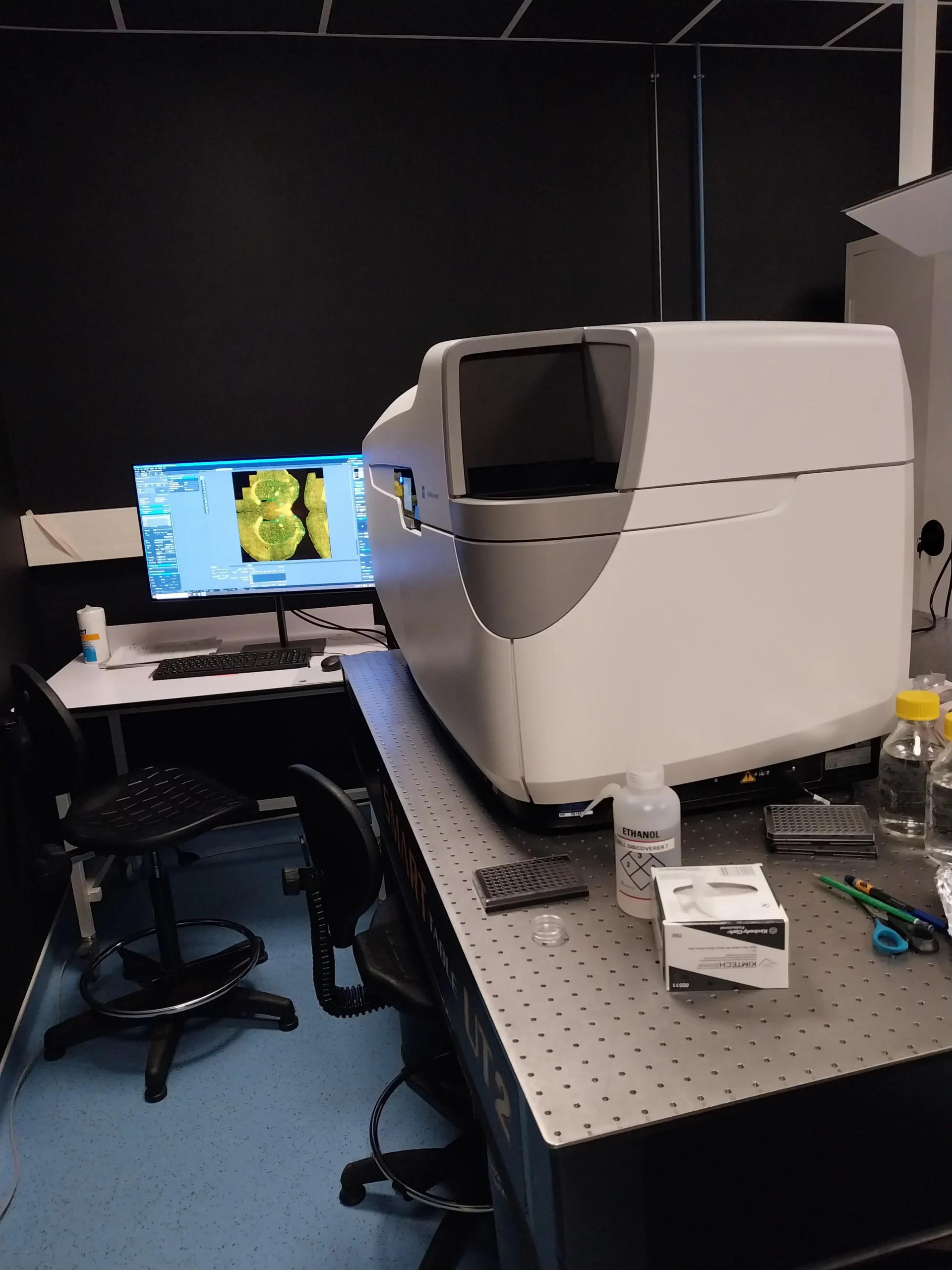
Zeiss Cell Discoverer 7
Specifically dedicated to High-content imaging and screening, this microscope is a medium throughput automated wild-field microscope from Zeiss. All regular samples can be imaged, from slides to petri dishes and multi-well-plates: contact us for more information.
It is equipped with a Zeiss Axiocam 512 camera, 4 diodes Led (405nm, 488nm, 561nm, and 640nm) and a filter wheel for imaging fluorophores such as DAPI, GFP/ Alexa 488, mCherry/ Alexa 555 and Alexa 647. The available objectives are: a 5x/0.35 (dry), a 20x/0.7 (dry, long working distance) and a 50x/1.2 (water). Intermediate lenses (0.5x, 1x, or 2x) are used to adapt the sampling to the size of the detectors. It is, also, equipped with an environmental control, temperature/CO2/humidity for live samples.
The system is also equipped with an analysis tools that enables conditional acquisitions ( « Smart or Adaptive Feedback Microscopy ») : contact us for more information.
A dedicated, offline analysis station, equipped with Arivis and Zen Desk software is available for data processing.
Project 1
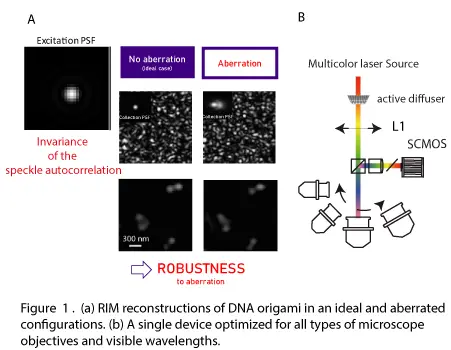
Despite their high price, super-resolved fluorescence microscopes, either scanning or structured illumination microscopes (SIM), used in biology imaging platforms, often show degraded performances due to sample induced optical aberrations.
We have recently developed an easy to use technique that provides the same resolution as SIM while being robust to aberrations. The Random Illumination Microscope (RIM) is based on the use of random dynamic illumination and statistical data processing. RIM has demonstrated its ability to image at high resolution samples that were inaccessible to current super-resolution techniques [Mangeat2021].
The objective of this project is to realize a fibered dynamic random illumination module that can be adapted to all microscopes. By accompanying this module with an adapted data processing algorithm, super-resolved microscopy will become accessible to all biology laboratories at a lower cost (less than 20 000 euros).
This project is a partnership between the CBI, the Fresnel Institute and the French SME Oxxius specialized in the realization of laser sources in health biology.
Project 2
The major challenge in cell and developmental biology is to propose a multi-scale model linking the action of macromolecules to cell function and then to the remodelling of biological tissue. One major application is to understand how stress induced by mechanical forces or DNA damage leads to cancer. Studying this requires observations at different scales (from sub-100 nm to mm), which are currently provided by different instruments, making it difficult to gain a global, integrated understanding of the process. A microscope capable of imaging the dance of proteins in large volumes of cell tissue would be a major asset for the development of integrated models in biology.
Recently, we have developed a random illumination microscope (RIM), which combines the resolution of the best SIMs with the ease of use and applicability of the standard fluorescence microscope. RIM involves illuminating the sample with a series of random ‘speckles’ and reconstructing a super-resolved image from the second-order statistics of the data. It is based on a mathematical analysis proving that the resolution of such a system can be equivalent to that of SIM. As the speckle statistic is insensitive to aberrations, RIM should work in conditions that SIM cannot. Over the last two years, RIM has been implemented in a simplified version in which the sample to be reconstructed is considered as a thin slice restricted to the focal plane (2D-RIM). Its performance already positions it as one of the best super-resolved optical microscopy techniques for studying living organisms (a resolution of 120 nm transverse and 300 nm axial at a rate of 1-5 Hz). However, it is clear that 2D-RIM does not exploit all the possibilities of RIM. In particular, taking into account the three-dimensional structuring of the speckle and the microscope’s impulse response in the data analysis should make it possible to significantly improve the axial and temporal resolution.
In this project, we propose to extend the principle of RIM into three dimensions with appropriate mathematical analysis and data processing, coupled with improved instrumentation. Our aim is to provide images over a large field of view with a transverse resolution of 100 nm and an axial resolution of 200 nm, and to achieve an image rate of 10 to 30 Hz. 3D-RIM will be used to answer two major biological questions for which no current super-resolution microscope is operational: the interaction of cells with the surrounding tissue during apoptosis, which requires coupling high spatial resolution to large fields of view, and the dynamic extrusion of the chromatin loop during DNA repair, which requires high spatiotemporal resolution.
Project 3
The spatial organization of DNA in the nucleus plays a key role in the mechanisms of gene expression regulation. In metazoan cells, the DNA fiber is organized around histone molecules to form the 30nm chromatin fiber. This fiber folds back on itself and has higher levels of organization known to be involved in the regulation of gene expression. This organization is nowadays mainly studied by chromatin contact maps methods. This is a statistical approach that does not allow us to explain the mechanisms at the scale of the single cell. We aim to establish, via imaging techniques, the structure of chromatin in the nucleus as well as the organization, relative to chromatin, of different proteins in the nucleus involved in gene regulation.
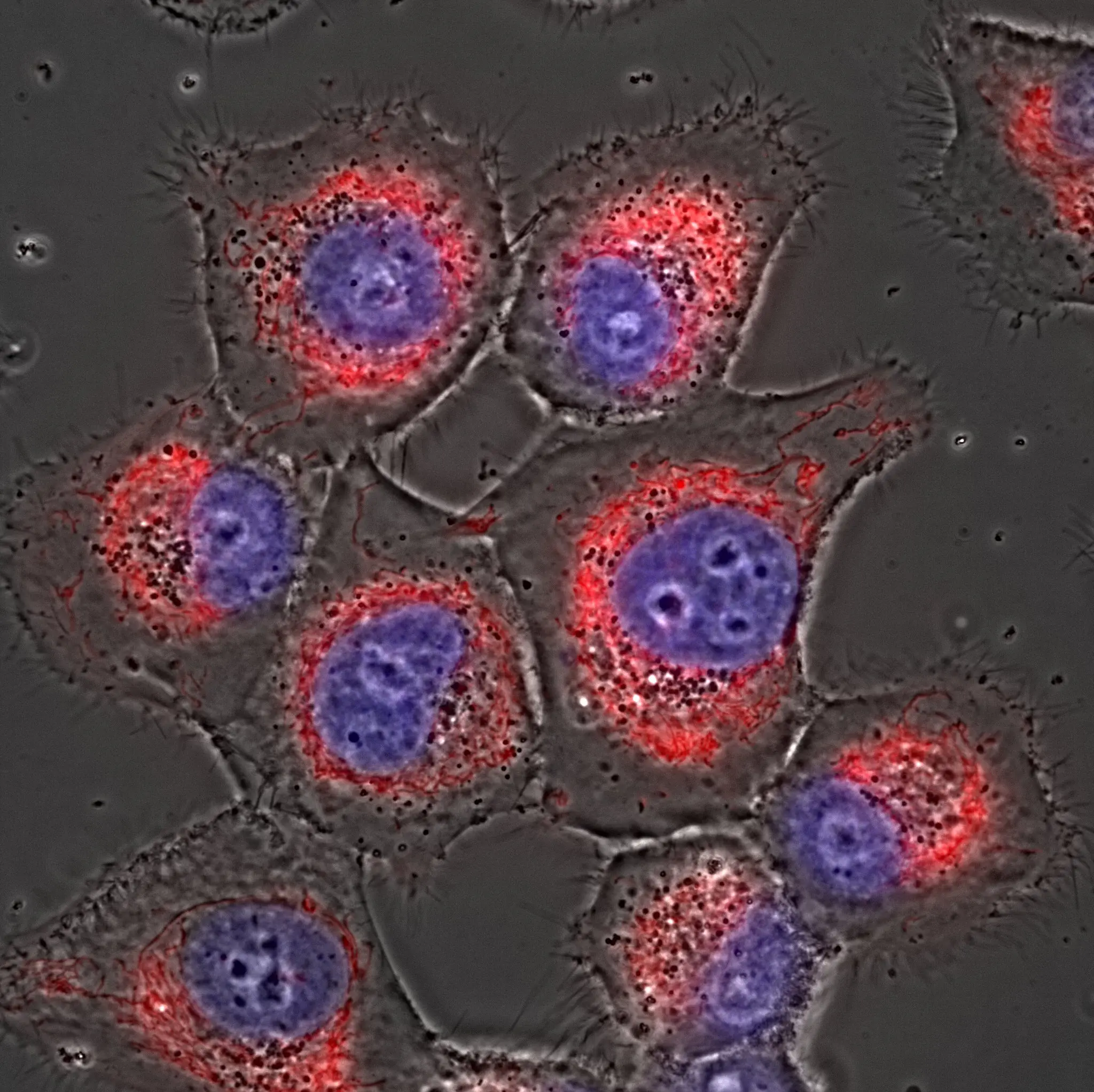
The chromatin fiber and the various proteins that interact with it form very dense structures that are particularly difficult to image. In the literature, imaging of chromatin by structured illumination microscopy techniques shows the existence of domains. These domains have the size of the resolution limit (around 100 nm). These technics does not allow to determine the structure underlying these domains. However, we are able to perform this type of chromatin live imaging thanks to the RIM (Random Illumination Microscopy) system developed on the platform with an excellent spatiotemporal resolution.
In order to go further, we are studying the structure of chromatin via SMLM (Single Molecule Localization Microscopy) approaches, which theoretically allow access to higher resolution. We apply these techniques using the yeast S. cerevisiae as a model. Indeed, its genome is smaller and less condensed than in other more complex organisms. However, the density of the structures studied remains very high and it is a challenge for SMLM microscopy, which requires both the use of a very stable imaging system and a perfect mastery of the signal processing approaches inherent to this technology. We therefore need a high-performance SMLM system, allowing high acquisition rates while integrating signal processing approaches (variable blurs, deconvolution in the Zernike base). We are also developing an original approach that combines DNA imaging (DNA-PAINT) and PALM microscopy of chromatin partner molecules. For this, we need an imaging system that allows us to perform simultaneous two-color SMLM acquisition. Engineers of the platform have already acquired a great expertise in the different aspects of this project.
– Guillaume Giroussens, Simon Labouesse, Marc Allain, Thomas Mangeat, Lorry Mazzella, Loıc le Goff, Anne Sentenac, Jerome Idier. Fast super-resolved reconstructions in fluorescence random illumination microscopy (RIM. IEEE TCI 2024 Dec
– Lorry Mazzella, Thomas Mangeat, Guillaume Giroussens, Benoit Rogez, Hao Li, Justine Creff, Mehdi Saadaoui, Carla Martins, Ronan Bouzignac, Simon Labouesse, Jérome Idier, Frédéric Galland, Marc Allain, Anne Sentenac & Loïc LeGoff. Extended-depth of field random illumination microscopy, EDF-RIM, provides super-resolved projective imaging Light Science & Applications 2024 Oct
– Marco Paoli, Antoine Wystrach, Brice Ronsin, Martin Giurfa. Analysis of fast calcium dynamics of honey bee olfactory coding Elife 2024 Sep
– Simon Labouesse, Jérôme Idier, Marc Allain, Guillaume Giroussens, Thomas Mangeat, Anne Sentenac. Superresolution capacity of variance-based stochastic fluorescence microscopy: From random illumination microscopy to superresolved optical fluctuation imaging Physical Review A 2024 Mar
– Silvia Kocanova, Flavien Raynal, Isabelle Goiffon, Betul Akgol Oksuz, Davide Baú, Alain Kamgoué, Sylvain Cantaloube, Ye Zhan, Bryan Lajoie, Marc A Marti-Renom, Job Dekker, Kerstin Bystricky. Enhancer-driven 3D chromatin domain folding modulates transcription in human mammary tumor cells Life Sci Alliance 2023 Nov
– Marie Zilliox, Vanessa Tillement, Thomas Mangeat, Sophie Polès, Patrick Blader and Julie Batut. Protocol to locally express cxcl12a during zebrafish olfactory organ development by combining. IR-LEGO with live imaging. Star Protocols 2023 Aug
– Kévin Affannoukoué, Simon Labouesse, Guillaume Maire, Laurent Gallais, Julien Savatier, Marc Allain, Md Rasedujjaman, Loic Legoff, Jérôme Idier, Renaud Poincloux, Florence Pelletier, Christophe Leterrier, Thomas Mangeat, and Anne Sentenac. Super-resolved total internal reflection fluorescence microscopy using random illuminations. OPTICA 2023 Jul
– Jasnin M, Hervy J, Balor S, Bouissou A, Proag A, Voituriez R, Schneider J, Mangeat T, Maridonneau-Parini I, Baumeister W, Dmitrieff S, Poincloux R. Elasticity of podosome actin networks produces nanonewton protrusive forces Nature Communication 2022 Jul
– Marion Portes, Thomas Mangeat, Natacha Escallier, Ophélie Dufrancais, Brigitte Raynaud-Messina, Christophe Thibault, Isabelle Maridonneau-Parini, Christel Vérollet and Renaud Poincloux. Nanoscale architecture and coordination of actin cores within the sealing zone of human osteoclasts eLife 2022 Jun
– Emilie L Cerezo, Thibault Houles, Oriane Lié, Marie-Kerguelen Sarthou, Charlotte Audoynaud, Geneviève Lavoie, Maral Halladjian, Sylvain Cantaloube, Carine Froment, Odile Burlet-Schiltz, Yves Henry, Philippe P Roux, Anthony K Henras, Yves Romeo. RIOK2 phosphorylation by RSK promotes synthesis of the human small ribosomal subunit PLoS Genet 2021 Jun
– Nicolas Giang, Marion Mars, Marc Moreau, Jose E Mejia, Grégory Bouchaud, Antoine Magnan, Marine Michelet, Brice Ronsin, Geoffrey G Murphy, Joerg Striessnig, Jean-Charles Guéry, Lucette Pelletier, Magali Savignac. Separation of the CaV 1.2-CaV 1.3 calcium channel duo prevents type 2 allergic airway inflammation Allergy 2022 Feb
– Thomas Mangeat, Simon Labouesse, Marc Allain, Awoke Negash, Emmanuel Martin, Aude Guénolé, Renaud Poincloux, Claire Estibal, Anaïs Bouissou, Sylvain Cantaloube, Elodie Vega, Tong Li, Christian Rouvière, Sophie Allart, Debora Keller, Valentin Debarnot , Xia Bo Wang , Grégoire Michaux, Mathieu Pinot, Roland Le Borgne, Sylvie Tournier, Magali Suzanne, Jérome Idier, Anne Sentenac. Super-resolved live-cell imaging using random illumination microscopy Cell Rep Methods 2021 April
– Noémie Kempf, Fatima Moutahir, Isabelle Goiffon, Sylvain Cantaloube, Kerstin Bystricky, Anne-Claire Lavigne. Analysis of Cellular EMT States Using Molecular Biology and High Resolution FISH Labeling Methods Mol Biol 2021
Affiliation