team
RIBONOVA
Team leader: Gleizes Pierre-Emmanuel
Presentation
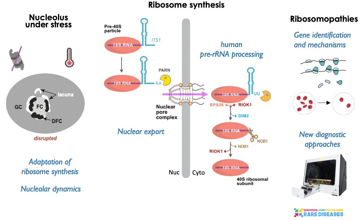
Ribosomes perform protein synthesis in all living organisms. They are present in abundance (5 to 10 million per human cell) and their own synthesis accounts for a significant proportion of the cell’s energy expenditure. The production of ribosomes and their activity in translation constitute a complex and dynamic system of mechanisms at the heart of gene expression and cellular metabolism. This system is subject to stringent regulation, adapting to the cell’s state of proliferation or differentiation, and responding to various stresses. Dysfunction of ribosome production and/or activity is at the root of a growing number of rare genetic diseases, called ribosomopathies, which affect various tissues. Our team focuses on the mechanisms of ribosome formation in human cells. Using a multifaceted approach encompassing molecular, cellular, and structural biology, we seek to elucidate the intricacies of pre-ribosome maturation into ribosomal subunits and to comprehend the dynamics of the nucleolus, the nuclear domain linked to ribosome formation. In parallel, we investigate the disruption of ribosome synthesis in ribosomopathies, particularly Diamond-Blackfan anemia, to elucidate pathophysiological mechanisms and enhance diagnostic capabilities. These studies are carried out in close collaboration with geneticists and clinicians worldwide. Our research also explores the involvement of the ribosome synthesis in the stress response.
Project 1
Ribosomal RNAs constitute the scaffold of the ribosomal subunits. Three of the four ribosomal RNAs (rRNAs) are embedded in a common precursor encoded in the rDNA. Processing of this precursor gradually tailors the mature rRNA by removal of the so-called transcribed spacers. Pre-rRNA processing is coordinated with the folding of the RNA and the assembly of the ribosomal proteins, which is orchestrated by a large number of ribosome biogenesis factors. While most of ribosome biogenesis takes place in the nucleus, and more specifically in the nucleolus, maturation of pre-ribosomal particles is only completed after export to the cytoplasm.
– Roles of 3′ exonucleases in human pre-rRNA processing. We and others have shown specific roles of 3′ exonucleases in mammalian pre-rRNA processing. Hence, we identified the poly-A specific ribonuclease (PARN) as the enzyme that ensures exonucleolytic trimming of the 3’ end of the 18S-E pre-rRNA, the last precursor to the 18S rRNA (Montellese et al., NAR 2017; collaboration: U. Kutay, ETH Zürich, A. Henras CBI). We are currently characterizing other exonucleases involved at various steps of human pre-rRNA processing and how 3′ adenylation or uridylation contributes to this process (PhD thesis of Maxime Aubert; collaboration: D. Gagliardi & H. Zubert, IBMP Strasbourg).
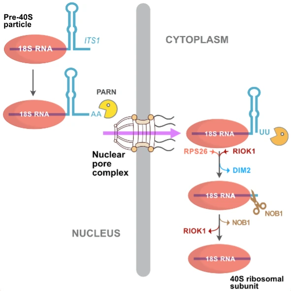
The late steps of human pre-40S particle maturation illustrate the role of exonucleases in pre-rRNA processing
– Control of the final cleavage at the 18S rRNA 3’ end by endonuclease NOB1 in the cytoplasm. The maturation of the 18S rRNA 3’ end occurs in the cytoplasm and completes the formation of 40S subunits. Using cryo-electron microscopy, we solved the structure of human pre-40S particles during late maturation stages and proposed a model where endonucleolytic cleavage by NOB1 is regulated by RBFs RIOK1, DIM2, and the addition of ribosomal protein RPS26. This model integrates in vitro and in cellulo experiments. (PhD thesis of Laura Plassart; Plassart et al. eLife 2022; collaboration: U. Kutay, ETH Zürich)
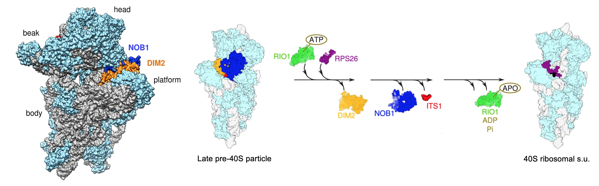
Adapted from Plassart et al, eLife, 2022
– Translocation of pre-ribosomal particles through the nuclear pore complex. Pre-ribosomal particles are abundant ribonucleoproteins crossing the nuclear pore complex (NPC). Using ultrafast freezing and electron tomography in yeast, we captured 3D snapshots of pre-ribosomal particles during nuclear export. We could then analyze their trajectory within the NPC and infer dynamic parameters of pre-ribosome nuclear export using a probabilistic model. (Delavoie et al. Nat. communication 2019; collaboration with Jean-Yves Dauxois, Toulouse Institute of Mathematics). We are currently analysing yeast strains with defective nucleoporins to further substantiate our model of pre-ribosomes’ translocation through the NPC.
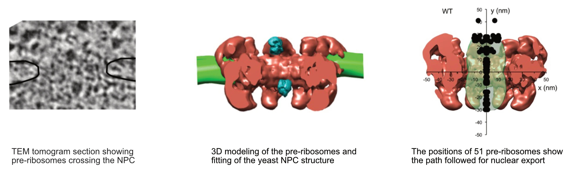
Adapted from Delavoie et al, Nature Communications, 2019
Project 2
Most ribosomopathies are congenital disorders associated with defects in ribosome synthesis. The Diamond-Blackfan anemia syndrome (DBAS) is a prototypical ribosomopathy in which patients usually present with bone marrow failure leading to a strong deficit of red blood cell production, as well as a variety of other developmental abnormalities. Over 95% of the cases with a genetic diagnosis show haploinsufficiency of a ribosomal protein gene, of which 24 have been linked to the disease so far. Several pathophysiological mechanisms have been proposed, including impaired translation due to a lower rate of cytoplasmic ribosomes or of aberrant ribosomes, nucleolar stress, dysregulation of heme metabolism. However, the tissue specificity of this disease still requires a definitive explanation. Of note, the genetic lesion remains unknown in 20-30% of the patients after genome sequencing. This significant challenge, which is shared with other rare diseases, calls for greater functional characterization in addition to high-throughput sequencing.
– New variants in ribosome biogenesis genes linked to ribosomopathies. We have long been collaborating with geneticists worldwide to characterize gene variants involved in DBA by studying their impact on ribosome synthesis. We recently identified deleterious variants in RPL8 and RPL17 (Lebaron et al, Human Mutation 2022; Fellmann et al., JCI Insight 2024), with RPL17 variants linked to atypical 60S ribosomal subunits containing a trimmed 5.8S rRNA. As a part of the European RiboEurope consortium (EJPRD), we also identified mutations in HEATR3, a ribosomal protein chaperone gene, in a new recessive form of the disease associated with intellectual disability (O’Donohue et al, Blood 2022).
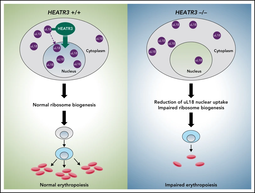
Adapted from O’Donohue et al, Blood, 2022
– Ribosome biogenesis defects in telomeropathies. In collaboration with Patrick Revy’s team (Imagine, Paris), we studied mutations in the NHP2 and PARN genes in patients with Høyeraal-Hreidarsson syndrome (a severe form of dyskeratosis congenita) or pulmonary fibrosis. Alteration of both genes is expected to affect the telomerase, since NHP2 is a component of the telomerase particle, and PARN an exonuclease involved in the maturation of TERC, the telomerase RNA. We found that, in addition to telomere dysfunction, these mutations caused defects in ribosome biogenesis, consistent with their known role in snoRNP formation and pre-rRNA maturation, respectively. This defect in ribosome synthesis may contribute to the pathogenic mechanisms and worsen the disease (Benyelles et al., EMBO Mol Med 2019; Benyelles et al., Hum Mol Genet 2020).
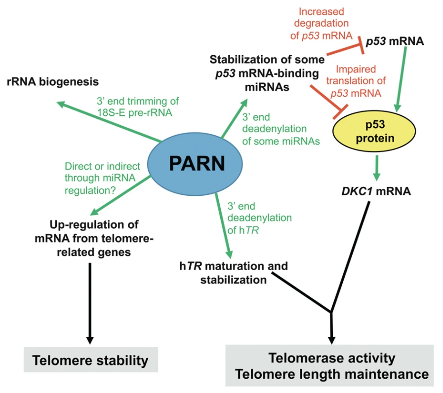
Adapted from Benyelle et al, EMBO Mol Med, 2020
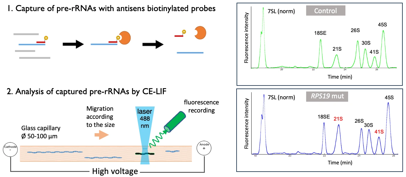
– New method to analyze pre-rRNAs by capillary electrophoresis for DBAS diagnosis. Improving the diagnosis of DBAS is crucial to avoid diagnostic wandering and provide the best care possible for the patients. We have developed a protocol using capillary electrophoresis coupled with laser-induced fluorescence (CE-LIF) for detecting pre-rRNA processing anomalies. This method demonstrated over 85% accuracy in distinguishing DBAS patients from controls and is being adapted for automation and standardization through a commercial solution in order to spread the technique to non-specialized laboratories. This work has been performed in close contact with WinSep and and Adelis, two companies based in Toulouse.
Project 3
Most of the ribosome biogenesis steps in eukaryotes take place in the nucleolus, a nuclear domain that organizes around the rDNA genes. In mammalian cells, the nucleolus adopts a tripartite structure that reflects the centrifuge movement of pre-ribosomes from the rDNA to the nuclear pore complex during their maturation. Auto-organization of this structure results from the condensation of its components, driven both by functional protein-protein, protein-RNA, or RNA-RNA interactions linked to ribosome biogenesis transactions and by spontaneous interactions of intrinsically disordered domains found in many nucleolar proteins, leading to liquid-liquid phase separation. Under certain stress, like heat-shock or proteotoxic stress, the nucleolus reorganises in conjunction with ribosome biogenesis interruption and formation of protein aggregates. We are currently collaborating with the team led by Dimitris Xirodimas (CRBM, Montpellier) to understand the mechanisms leading to the recovery of the nucleolus after a proteotoxic stress, in particular the role of the small IGS non-coding RNAs in this process. We managed to observe the protein aggregates and their gradual disappearance upon alleviation of the stress through high-resolution fluorescence microscopy and correlative light-electron microscopy, in collaboration with the CBI light microscopy and electron microscopy facilities (LITC, METi). Elimination of the aggregates involves the nuclear proteasomes as well the lncRNA IGS42, which was detected in the aggregates. This study shows that a protein quality control system operates in the nucleolus during stress recovery to eliminate these stress related-inclusions (Brunello et al., EMBO J. 2025).
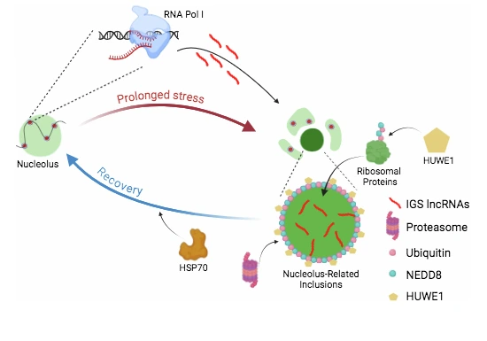
From Brunello et al. EMBO J., 2025
Project 4
Our team has long been involved in the implementation of new electron microscopy techniques, including electron tomography, correlative light-electron microscopy and cryo-EM. We recently developed a joint interdisciplinary research project with Etienne Snoeck at the CEMES in Toulouse, a CNRS laboratory specialized in material science. This laboratory has developed a unique 300 keV electron microscope to perform electron holography, a spectroscopic technique allowing recording of phase images and detection of electrostatic potentials in samples at nanometric scales. Together with the multiscale electron imaging facility of the CBI (METi), we have combined our expertise to explore the potential of this instrument to image biological samples at room temperature or in cryo mode without any staining. Images of T4 and T5 phages were obtained in off-axis or in-line mode revealing both promising capabilities in the measurement of local electrostatic potentials and important bottlenecks that will need to be addressed in the future. (PhD thesis of Elio Karim; Karim et al. J. Struct. Biol. 2025; collaboration: METi)
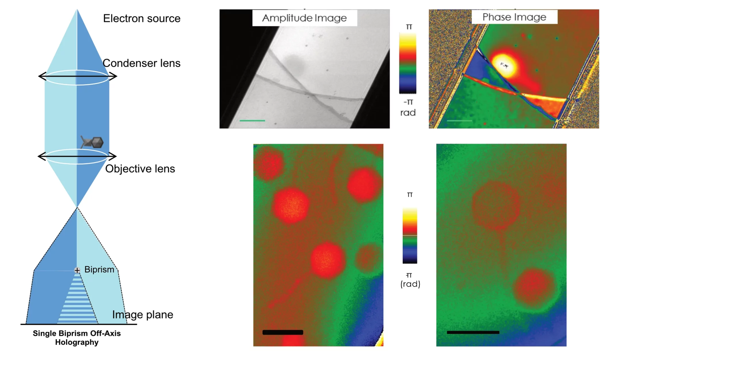
Adapté de Karim et al., J. Structural Biology, 2025
Team members
– Delavoie F et al. (2019) The path of pre-ribosomes through the nuclear pore complex revealed by electron tomography. Nat. Comm. 10:497
– Plassart L et al. (2021) The final step of 40S ribosomal subunit maturation is controlled by a dual key lock. eLife 10:e6154
– O’Donohue MF et al. (2022) HEATR3 variants impair nuclear import of uL18 (RPL5) and drives a Diamond-Blackfan anemia syndrome in humans. Blood 139:3111-26
– Fellman F, Saunders C, O’Donohue MF et al. (2024) An atypical form of 60S ribosomal subunit in Diamond-Blackfan anemia linked to RPL17 variants. JCI Insight, 9:e172475
– Brunello L et al. (2024) A nuclear protein quality control system for the elimination of nucleolus-related inclusions. EMBO J, online ahead of print (https://doi-org.insb.bib.cnrs.fr/10.1038/s44318-024-00333-9)