Team
Team Leader: Bystricky Kerstin
Presentation
Do chromatin structure, chromosome motion and genome organization regulate nuclear processes? To address this question, we investigate the functional relationship between the location of a gene, its inherent dynamics and its response to stimuli. We analyze epigenetic mechanisms of gene expression in mammary tumor cells and fibroblasts, investigating in particular the role of histone variants and 3D folding of chromatin domains at genes sensitive to stimuli. We develop techniques to visualize and image DNA in living cells, at the level of single loci and the whole genome, at high resolution, and methods to quantitatively analyze and model the data. Our challenge is experimentally acting upon structure to probe impact on function.
Project 1
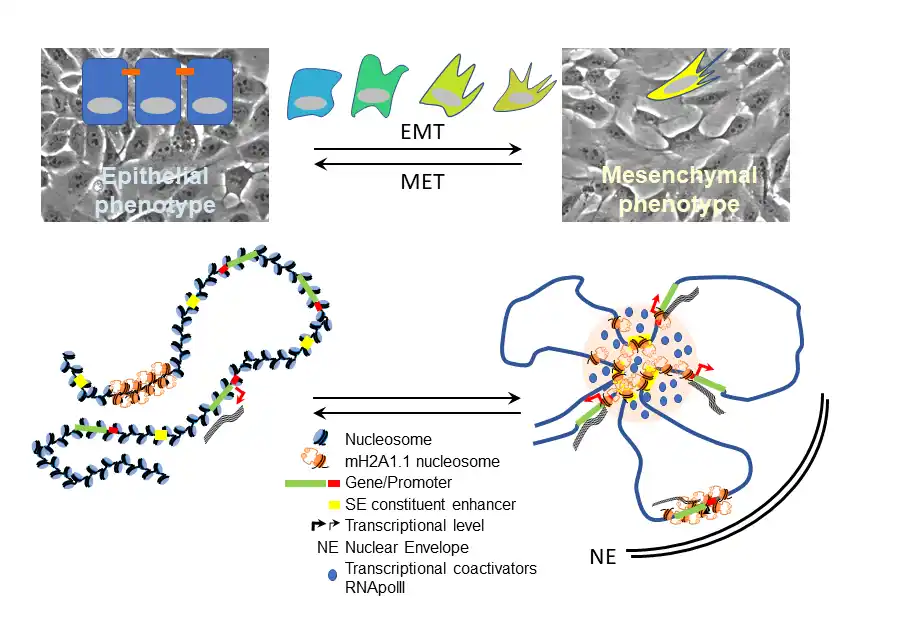
The principal aim of this project is to characterize the role of mH2A1.1 in cellular plasticity of TNBC cells by analyzing its effect on gene expression, 3D genome architecture and biophysical properties.
Unlike canonical histones, variants like mH2A1.1 have distinct structures, allowing them to influence chromatin architecture and gene expression. mH2A1.1 plays a role in critical cellular processes, such as epithelial-to-mesenchymal transition (EMT) and cell metabolism. We have shown that its overexpression is associated with poor prognosis in TNBC.
We are now focusing on characterizing the molecular mechanisms that enable mH2A1.1 to function as a transcriptional regulator in TNBC cells. Notably, we found that mH2A1.1 binds to super-enhancers (SEs), in addition to gene promoters. SEs are membraneless organelles that form through Liquid-Liquid Phase Separation (LLPS), a process driven by intrinsically disordered regions in transcription factors and coactivators. Given the disordered and highly charged nature of mH2A1.1’s linker domain, it is possible that it contributes to LLPS at SEs, amplifying gene expression.
Our goal is to identify the partners involved in its dual transcriptional activities, encompassing both its repressive and activating functions, as well as to explore its potential Liquid-Liquid Phase Separation (LLPS) properties, which may be key to its functional roles. At the cellular level, we are investigating the impact of mH2A1.1 on maintaining specific mesenchymal traits and the cellular plasticity of MDA-MB231 cells, and how this influences their propensity to develop metastasis.
Project 2
Chromatin organization in eukaryote cells has been extensively studied from nucleosome to genome, revealing the importance of chromatin spatial organization in regulating cellular functions. Temporal aspects of chromatin organization and its dynamic properties, on another hand, are often missing from analyses addressing regulation of DNA-related processes.
To study the involvement of chromatin dynamics in cellular processes, we developed complementary molecular and analysis tools. We are developing an original live-microscopy approach to describe and quantify chromatin dynamics at the scale of the entire nucleus called High resolution Diffusion mapping (HiD) in collaboration with the biophysicist team of Manoel Manghi at the LPT. HiD quantify local chromatin motion with sub-pixel accuracy to create two-dimensional maps of chromatin domain dynamics in single living cells. HiD can be combined with our original ANCHOR DNA labeling technology to analyse dynamics of a single locus in single cells and decipher the impact of transcription regulation on chromatin motion. We further apply genome wide approaches (HiC, ChIpSeq, micro C etc.) to study the epigenetic landscape in cells in response to pharmacological stimuli and/or mechanical stress in wild-type and genome edited cells. Using a combination of interdisciplinary approaches (HiD, polymer modelling and genomic data) we want to gain a mechanistical understanding of the link between 3D genome architecture, chromatin dynamic behavior and properties, and function.
An ongoing application of HiD involves studying the spatiotemporal chromatin dynamics during SAHF (senescence-associated heterochromatin foci) formation in oncogene-induced senescence (OIS) in human fibroblasts. This includes investigating the role of epigenetic SAHF biomarkers such as macroH2A1 and histone post-translational modifications. Additionally, we are exploring the properties of certain small natural molecules that act on chromatin. Recently, we identified a new set of plant-derived molecules capable of inducing SAHF formation, as well as others that trigger light-induced chromatin modifications.
Team members
– Kocanova S., Raynal F., Goiffon I, Oksuz BA, Baú D, Kamgoué A, Cantaloube S, Zhan Y, Lajoie B, Marti-Renom MA, Dekker J, Bystricky K “Enhancer-driven local 3D chromatin domain folding modulates transcription in human mammary tumor cells” Life Science Alliance, 7(2), 2024
– Recoules, L., Tanguy Le Gac, N., Moutahir, F., Bystricky, K. & Lavigne, A. C#. (2023). Recruitment of the Histone Variant MacroH2A1 to the Pericentric Region Occurs upon Chromatin Relaxation and Is Responsible for Major Satellite Transcriptional Regulation. Cells 12. https://doi.org/10.3390/cells12172175
– Recoules, L., Heurteau, A., Raynal, F., Karasu, N., Moutahir, F., Bejjani, F., Jariel-Encontre, I., Cuvier, O., Sexton, T., Lavigne, A.C#, and Bystricky K#. (2022). The histone variant macroH2A1.1 regulates RNA polymerase II-paused genes within defined chromatin interaction landscapes. Journal of Cell science 135. https://doi.org/10.1242/jcs.259456
– Kempf, N., Moutahir, F., Goiffon, I., Cantaloube, S., Bystricky, K., and Lavigne, A.C#. (2021). Analysis of Cellular EMT States Using Molecular Biology and High Resolution FISH Labeling. Methods Mol Biol 2179, 353-383. https://doi.org/10.1007/978-1-0716-0779-4_27
– Barth R., Bystricky K. and Shaban HA “Coupling chromatin structure and dynamics by live super-resolution imaging” Science Advances 6, 2020
– Shaban HA, Barth R., Recoules, L. and Bystricky K. “HiD: Nanoscale mapping of nuclear dynamics in living human cells”. Genome Biology 21(1), 2020
– Germier T, Kocanova S, Walther N, Bancaud A, Shaban HA, Sellou H, Politi AZ, Ellenberg J, Gallardo F, Bystricky K (2017) Real-Time Imaging of a Single Gene Reveals Transcription-Initiated Local Confinement. Biophysical journal 113: 1383-1394
– Lavigne AC, Castells M, Mermet J, Kocanova S, Dalvai M, and Bystricky K. Increased macroH2A1.1 Expression Correlates with Poor Survival of Triple-Negative Breast Cancer Patients. PLoS ONE, 2014, 9 (6), pp.e98930. https://doi.org/10.1371/journal.pone.0098930
Affiliation