Team
Team leader: Wang Xiaobo
Presentation
Cell migration is crucial for development and cancer. Migrating cells display different migratory behaviors and chemical-mechanical responses when they encounter different microenvironments including matrix and substrate cells. Thus, an important question is how migrating cells interact with and respond to different microenvironments. In addition, cell migration is often integrated with other cellular processes such as cell proliferation. How cell migration is integrated with cell proliferation is a key to understanding cancer initiation and progression.
The team uses a complementary set of molecular, genetic and imaging strategies and mathematical and physical modeling to study 1) how different types of invasive cell migration occur, are controlled and play a role in tissue morphogenesis and cancer progression, and 2) how invasive cell migration is linked and integrated with cell division during cancer progression. The team is experienced in many imaging techniques (FRET biosensors, optogenetics, RIM super-resolution microscopy, and expansion microscopy) and some biophysics tools (laser ablation, optical tweezer).
Project 1
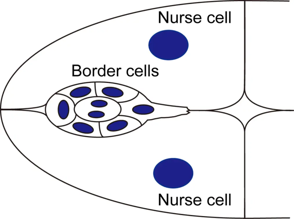
Collective cell migration during development and pathological processes often occurs in the cell-rich tissues which are composed of not only matrix but also substrate cells. The characteristics and role of substrate cell environment are poorly understood, compared with the ones of matrix environment. The team uses Drosophila border cell migration as an in vivo model to understand how collective cells interact and communicate with substrate cells to pass through a cell-rich tissue.
Our previous study identified actomyosin pulses as a critical factor controlling border cell migration (Combedazou et al, J Cell Sci, 2017). We also revealed two functional Rac1 pools at border cell protrusions and cables, and the balance of contractile and protrusive Rac1 pools can integrate the direction and coordination of border cell migration (Zhou et al, Nat Commun, 2022). Regarding migrating border cells, we will explore the origin, control and role of actomyosin pulses for migration efficiency. In addition, we lately revealed that Rac1 is also critical for substrate cell environment for the travel path, thus affecting migration efficiency. Regarding substrate cell environment, we will explore the key factors present in substrate cells that control border cell migration behavior and efficiency, which is little explored. Understanding substrate cell environment will facilitate us to better know how cancer cells migrate through similar stroma cell environment during cancer invasion and metastasis.
Project 2
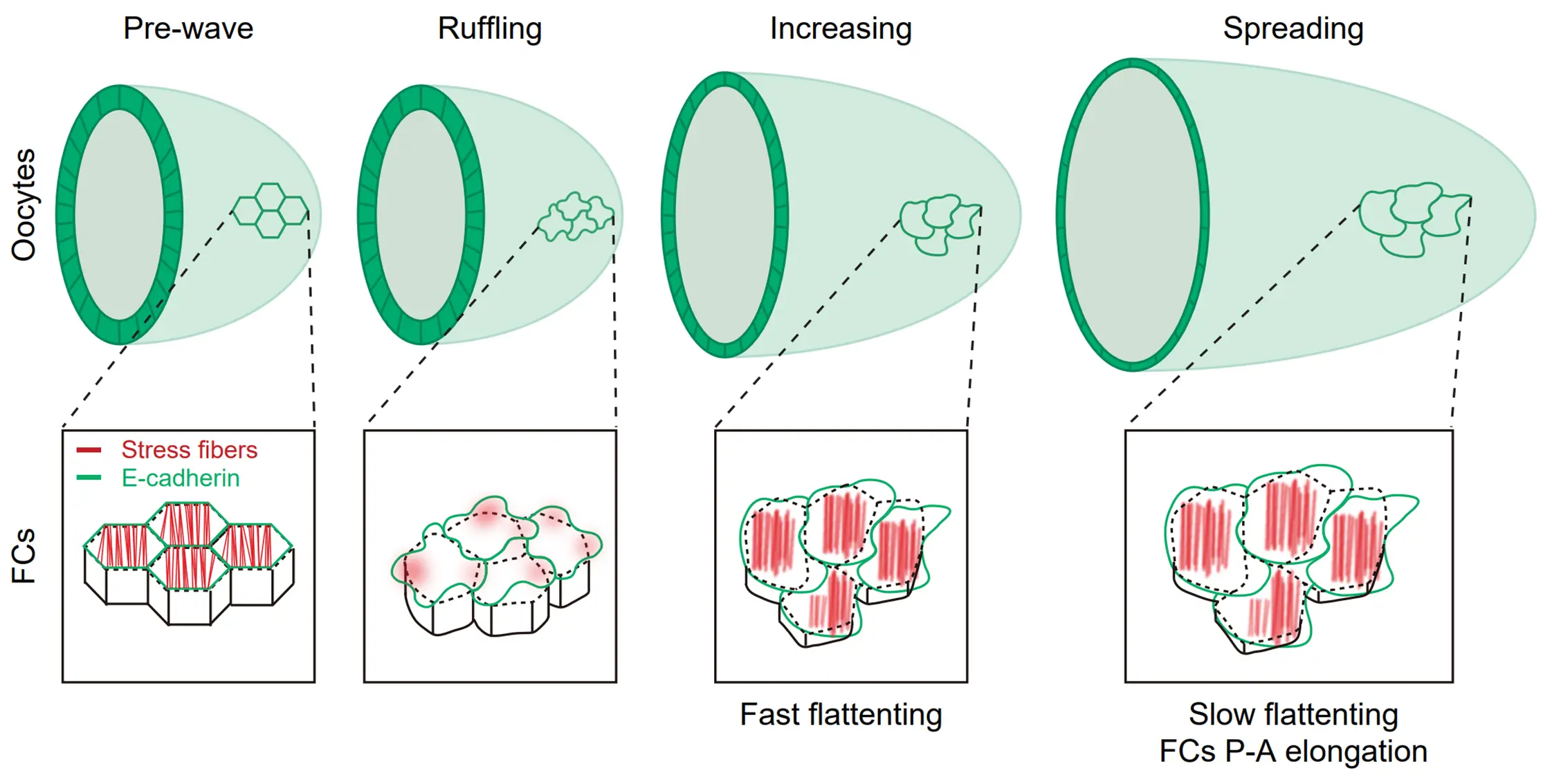
Stress fibers are one of the important cytoskeleton networks by which epithelial cells interact and communicate with extracellular matrix, and thus play various important roles in controlling cell proliferation, migration and homeostasis. Compared with cultured cells, our understanding of stress fibers in vivo is very limited. The team uses Drosophila follicle epithelial cells as an in vivo model to understand the origin, modulation, mechanics and function of basal stress fibers in epithelial cells in vivo.
Our previous study provided some mechanistic insights into contractile basal stress fibers: 1) interaction with different adhesions (Qin et al, Nat Commun, 2017); 2) controlling mechanism of basal myosin pulses (Qin et al, Nat Commun, 2018); 3) supracellular cytoskeleton network (Popkova et al, Nat Commun, 2020).
Regarding these contractile stress fibers, we will explore two main questions: 1) the control of basal F-actin networks and pulses, and 2) the origin and propagation of basal actomyosin pulses. In addition, we lately unraveled expanding basal stress fibers that control a novel epithelial expansion behavior to integrate cell flattening and oocyte elongation (Li et al, Nat Commun, 2022).
Regarding these expanding stress fibers, we will explore three main questions: 1) the switch from contractile to expanding stress fibers; 2) the mechanics of expanding stress fibers; 3) the correlation between expanding stress fibers and tissue adaptation. Understanding different stress fibers in vivo will facilitate us to better know how epithelial cells remodel their cytoskeleton networks to modulate their different processes during development and cancer progression.
Project 3
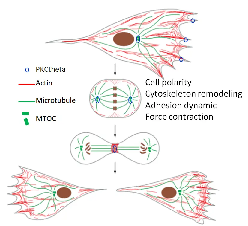
Cancer is caused by the deregulation of fundamental biological processes in normal cells. Although both abilities of cell migration and cell division are often gradually enhanced during cancer development and progression from non-invasive to highly metastatic types, the link between these two cellular behaviors are seldom explored. Here an important question is whether and how these two cellular behaviors are integrated together by some common controls. Using highly metastatic breast and ovarian cancers as a model to study tumor aggressiveness, we found that the serine/threonine kinase PKCtheta, a key enzyme in T lymphocyte activation, is highly expressed in these particularly aggressive subtypes of cancer (Chadelle et al, Cancer Letters 2022). PKCtheta presents several specific subcellular localizations in these aggressive cancer cells implicating various biological functions. Indeed, PKCtheta is involved in the control of proliferation, migration and invasion of aggressive cancer cells as well as their metastatic activity. Here, our main objective is to determine whether and how PKCtheta uses common factors and mechanisms such as cell polarity, cytoskeleton remodeling, adhesion dynamic and force contraction, to govern and coordinate cell migration and cell division. This integrated behavior would undoubtedly amplify the impact of PKCtheta on tumor aggressiveness.
– Li S, Liu Z-Y, Li H, Zhou S, Liu J, Sun N, Yang K-F, Dougados V, Mangeat T, Belguise K, Feng X-Q, Liu Y, Wang X*. Basal actomyosin pulses expand epithelium coordinating cell flattening and tissue elongation. Nat Commun (2024) 15(1):3000.
– Zhou S, Li P, Liu J, Liao J, Li H, Chen L, Li Z, Guo, Q, K Belguise, Yi B, Wang X*. Two Rac1 pools integrate the direction and coordination of collective cell migration. Nat Commun (2022) 12;13(1):6014.
– Chadelle L, Liu J, Choesmel-Cadamuro V, Karginov AV, Froment C, Burlet-Schiltz O, Gandarillas S, Barreira Y, Segura C, Van Den Berghe L, Czaplicki G, Van Acker N, Dalenc F, Franchet C, Hahn KM, Wang X*, Belguise K*. PKCθ-mediated serine/threonine phosphorylations of FAK govern adhesion and protrusion dynamics within the lamellipodia of migrating breast cancer cells. Cancer Lett (2022) 526:112-130.
– Popkova A, Stone OJ, Chen L, Qin X, Liu C, Liu J, Belguise K, Montell DJ, Hahn KM, Rauzi M, Wang X*. A Cdc42-mediated supracellular network drives polarized forces and Drosophila egg chamber extension. Nat Commun (2020) 11(1):1921.
– Wang H, Qiu Z, Xu Z, Chen SJ, Luo J, Wang X*, Chen J*. aPKC is a key polarity molecule coordinating the function of three distinct cell polarities during collective migration. Development (2018) 145(9).
– Qin X, Hannezo E, Liu J, Mangeat T, Choesmel-Cadamuro V, Macdonald J, Liu Y, Yi B, Wang X*. A biochemical network controlling basal myosin oscillation. Nat Commun (2018) 9(1):1210.
– Qin X, Park BO, Liu J, Chen B, Choesmel-Cadamuro V, Belguise K, Heo WD, Wang X*. Cell-matrix adhesion and cell-cell adhesion differentially control basal myosin oscillation and Drosophila egg chamber elongation. Nat Commun (2017) 8:14708.
– Combedazou A, Choesmel-Cadamuro V, Gay G, Liu J, Dupré L, Ramel D, Wang X*. Myosin II governs collective cell migration behaviour downstream of guidance receptor signalling. J Cell Sci (2016) pii: jcs.179952.
Affiliation