Team
Presentation
The transformation of a single fertilized egg into hundreds of thousands of cells that form the various organs of a developing embryo is a captivating process. To achieve this complex organization, embryonic cells simultaneously divide, arrange into tissues, and specialize into diverse cell types. While these processes follow specific temporal and spatial patterns, recent research has uncovered unexpected variability at the molecular, cellular, and tissue levels.
Our team aims to decipher the molecular and cellular rules governing embryonic self-organization and morphogenesis. We investigate how cell proliferation, migration, adhesion, and specialization are coordinated, with a focus on understanding how variability contributes to the plasticity and robustness of developmental processes.
We focus on the development of posterior tissues in vertebrate embryos, using birds (chickens and quails) as model systems. Our methods combine classical embryology techniques, such as grafting and electroporation, with time-lapse imaging, image analysis, and molecular approaches. Our findings have implications for various diseases, including congenital malformations and cancer. A key feature of our team is the establishment of multidisciplinary collaborations with mathematicians, physicists, and computer scientists, which is essential for addressing the complexity of biological systems.
Project 1
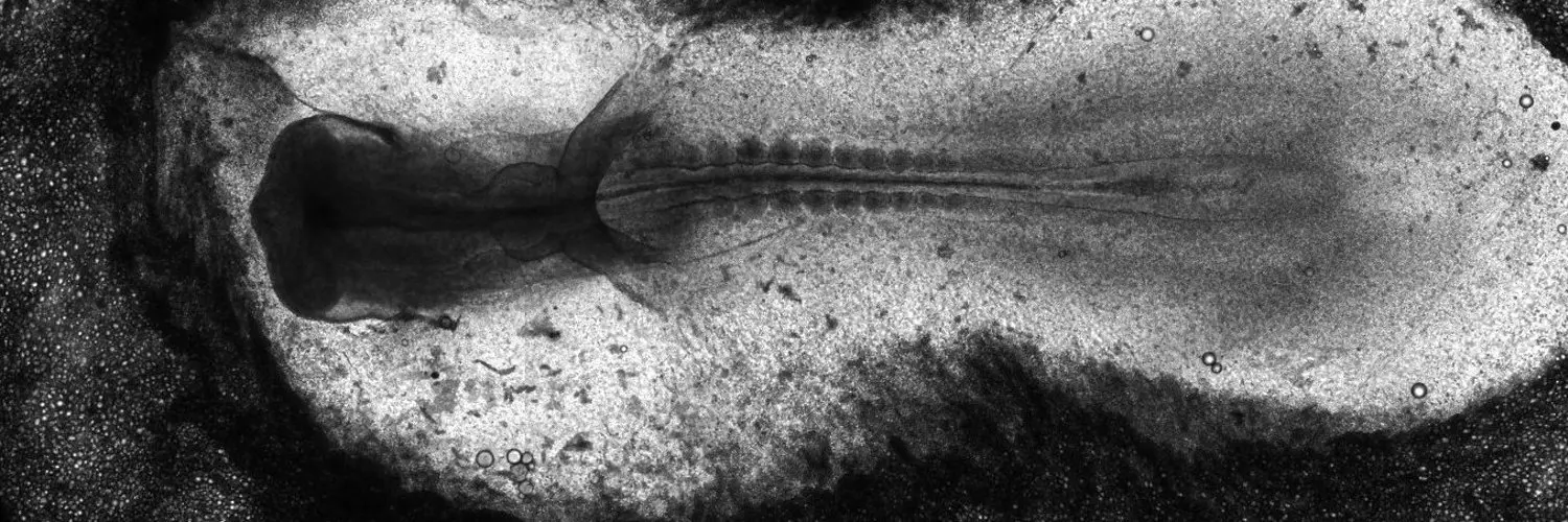
One of the key aspects of vertebrate embryo morphogenesis is the extension of its body along the head-to-tail axis. Through time-lapse imaging of transgenic quail embryos, we previously revealed that this body axis extension involves a complex choreography where ectodermal, mesodermal, and endodermal tissues proliferate, migrate, adhere differently, and slide past each other. Our current project aims to uncover the roles of these distinct cellular behaviors within the various tissues of the posterior embryonic body that drive this multi-tissue morphogenetic event. To achieve this, we use a combination of morphometric measurements, live imaging, functional studies, and modeling.
Project 2
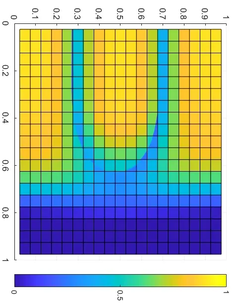
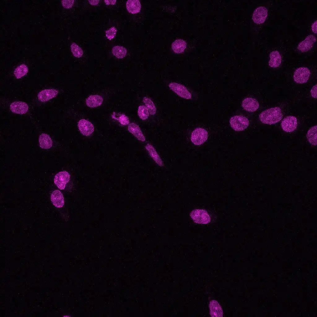
In the tail of growing vertebrate embryos, there is a region called the progenitor zone, which contains stem cells that give rise to mesodermal (future muscles and bones) and neural cells (future nervous system). As the embryo extends from head to tail, these cells self-renew and continuously supply the cellular material that will form the body’s blueprint. We discovered cell-to-cell variations in the expression levels of key transcription factors involved in progenitor fate specification within this cell population. These differences in expression levels influence whether stem cells remain in their niche or migrate into neural and mesodermal tissues. Mathematical modeling has shown that this cell-to-cell heterogeneity in the progenitor zone is a crucial factor in regulating cell rearrangement and tissue morphogenesis. Our current project aims to further understand how specification and morphogenetic programs are interconnected during axis extension.
– Romanos M, Salisbury T, Stephan S, Lansford R, Degond P, Trescases A, Bénazéraf B. Differential proliferation regulates multi-tissue morphogenesis during embryonic axial extension: integrating viscous modeling and experimental approaches. Development 2024 Jul
– Daniela Roellig, Sophie Theis, Amsha Proag, Guillaume Allio, Bertrand Bénazéraf, Jérôme Gros, Magali Suzanne. Force-generating apoptotic cells orchestrate avian neural tube bending Developmental Cell 2022 Mar
– Michèle Romanos, Guillaume Allio, Myriam Roussigné, Léa Combres, Nathalie Escalas, Cathy Soula, François Médevielle, Benjamin Steventon, Ariane Trescases, Bertrand Bénazéraf. Cell-to-cell heterogeneity in Sox2 and Bra expression guides progenitor motility and destiny eLife 2021 Oct
– Elena Gonzalez-Gobartt , José Blanco-Ameijeiras , Susana Usieto , Guillaume Allio , Bertrand Benazeraf, Elisa Martí. Cell intercalation driven by SMAD3 underlies secondary neural tube formation
Funding
Affiliation