How do a few cells take shape and organize themselves starting with the embryo’s first cell divisions? In a study of the early stages of development in the nematode Caenorhabditis elegans, Hervé Turlier, Matthieu Perez, Fabrice Delbary (MCD-CBI), and their colleagues show that it is now possible to reconstruct a true spatiotemporal map of the mechanical forces that organize cells.
From the very first stages of embryonic division, cells must organize themselves in a very precise manner to build a functional organism. This architecture does not depend solely on genetic programs or biochemical signals; it also results from mechanical forces generated at the cell surface and at cell interfaces.
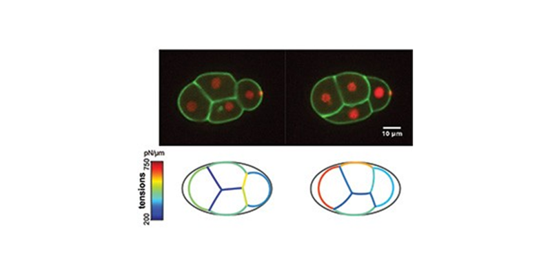
To make these forces measurable, scientists studied the early embryo of the nematode C. elegans at very early stages of development (2- and 4-cell stages), when the embryo exhibits a perfectly invariant cell lineage.
To shed light on the forces at play and to reconstruct their evolution during development, the study combines three complementary approaches: real-time microscopy images to track cell shape and measure the angles formed at their junctions; measurements taken using atomic force microscopy to obtain an absolute value for the stresses exerted on the cell surfaces; and, finally, a “foam” physical model to represent each cell as a bubble whose interfaces are subject to mechanical stresses. This approach has made it possible to create a spatiotemporal map of the forces that shape the embryo
Beyond the C. elegans model, this approach opens up new avenues for understanding how local changes in contractility or cell-cell adhesion result in changes in shape at the level of the entire embryo.
Latest news